Invisibility is cancer’s greatest weapon. Our MRI scan changes that.
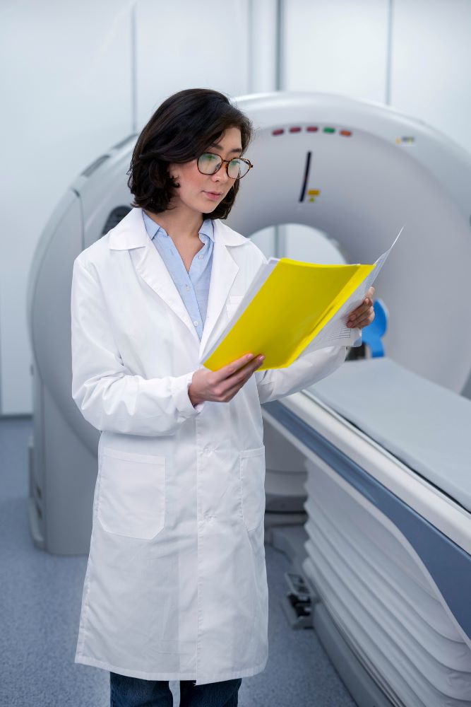
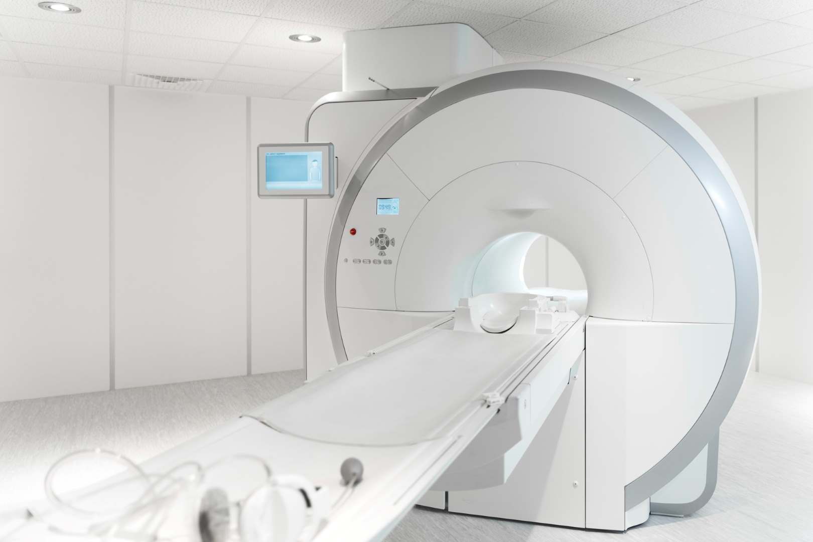
Magnetic Resonance Imaging (MRI) scans are uniquely effective at accurately displaying abnormalities inside the body. When combined with our world-class healthcare team, we make MRI-based full-body screening fast, accurate, and affordable.
WHY STAR MRI
Ultra-sensitive
Accurate
Fast*
Safe
Painless
*Our full body scan is 4x faster than a typical MRI scan.
WHAT WE SCAN
MRI, combined with our healthcare team and advanced technology, better empowers you to identify potential cancer than CT or ultrasound alone.
Useful screening tool
Not a useful screening tool
Useful, but utilizes harmful radiation
ORGAN
FULL BODY PLUS
CT
ULTRASOUND
Brain
Spine
Thyroid
Lung
Liver
Gallbladder
Pancreas
Spleen
Kidney
Adrenal Gland
Bladder
Ovary
Uterus
Prostate
Scroll to Top
Typical Symptoms
New onset or pattern change of headaches, unexplained nausea and vomiting, vision or hearing problems, difficulty balancing or walking, speech problems, personality or behavior changes, seizures.
Recent Research
MRI with AI Assisted Image Analysis: AI was used to assist radiologists in segmenting the brain into normal and abnormal tissue.
MRI for Brain Tumor Characterization: MRI was successfully used to differentiate high-grade and low-grade gliomas, as well as metastasis.
MRI for cancer screening systematic review: Evaluation of the Diagnostic Performance of Magnetic Resonance Spectroscopy in Brain Tumors: A Systematic Review and Meta-Analysis.
MRI Contrast: There was found to be no significant difference in the detection of brain tumors between contrast and non-contrast enhanced MRI scans in a cohort of children with prior brain tumors.
Typical Symptoms
Symptoms depend on tumor size, type, and location. Some common signs include pain, numbness, muscle weakness, difficulty standing or walking, spinal deformities, general loss of sensation and paralysis to varying degrees.
Recent Research
MRI vs. Other Imaging Modalities – MRI is considered the most reliable method for diagnosing spine tumors compared to CT and PET scans.
Typical Symptoms
Swelling, pain, or a lump in the front of the neck, trouble swallowing, a constant cough that is not due to a cold, changes in your voice.
Recent Research
MRI: MRI is highly accurate in differentiating benign and malignant lesions on the basis of apparent diffusion coefficient values obtained from diffusion weighted images.
Diffusion weighted MRI: DW-MRI images were used to risk stratify cases of thyroid cancer.
CT and MRI: CT and MRI were both found to be useful in assessing the extrathyroid extension of known cancer.
Typical Symptoms
Coughing up blood, shortness of breath, chest pain, losing weight without trying.
Recent Research
MRI: MRI can correctly identify malignant nodules 5mm or larger with increasing diagnostic accuracy when computer assisted detection is used.
Nodule Detection by MRI: MRI is suitable for lung cancer screening with excellent sensitivity and specificity for nodules greater than 6 mm.
CT and Chest X-Ray: This paper details the sensitivity and specificity of low-dose CT and chest X-Rays for lesion detection in a lung-cancer screening cohort.
MRI requires further study in order to determine efficacy in detecting lung cancer early with similar sensitivity and specificity as low dose computer tomography (LDCT). Therefore, the Ezra Full Body Scan includes a LDCT of the lungs for individuals at high risk of lung cancer, as determined by an Ezra medical provider.
Typical Symptoms
Most people do not show symptoms from early stage liver cancer. Symptoms may include losing weight without trying, loss of appetite, upper abdominal pain, nausea and vomiting, yellow discoloration of the skin/ eyes, white chalky stool.
Recent Research
MRI vs. CT and US – MRI was shown to be the best imaging modality for the detection of lesions in a high-risk population cohort when compared to CT or ultrasound.
Diagnostic Performance of MRI – MRI has a higher sensitivity and similar specificity for the detection of liver lesions when compared to CT and US.
Typical Symptoms
Abdominal pain/ bloating, fever, losing weight without trying, nausea, yellowing of the skin or eyes.
Recent Research
MRI vs. CT – MRI was found to be better than multidetector row CT in the diagnosis of gallbladder carcinoma.
Typical Symptoms
Signs and symptoms often don’t occur until the disease is advanced. They may include pain in the upper abdomen that radiates to your back, loss of appetite, unexplained weight loss, new-onset diabetes, yellowing of skin or eyes.
Recent Research
MRI vs. Other Imaging Modalities – In a large-population, mixed-patient cohort, MRI performed identically or better than CT, PET, or ultrasound for the detection pancreatic cancer.
Typical Symptoms
Enlarged spleen, pain in abdomen (upper left corner), unexplained weight loss, fever, fatigue, night sweats, weakness, and high levels of lymphocytes in the blood.
Recent Research
MRI: MRI is an excellent imaging tool for diagnosis, evaluation, and characterization of various focal splenic lesions and pathologic conditions.
Typical Symptoms
Signs and symptoms rarely present in the early stages of disease. In the later stages, kidney cancer may cause blood in urine, pain in the back/ side that doesn’t go away, loss of appetite, or unexplained weight loss.
Recent Research
MRI vs. CT – MRI showed similar sensitivity for the detection and staging of kidney cancer lesions as CT, and showed better specificity in both areas.
Non-Contrast MRI – Diffusion-weighted MRI can be used to effectively characterize renal lesions.
Typical Symptoms
Weight gain, muscle weakness, nausea, vomiting, back pain, fever, loss of appetite, pink or purple stretch marks on the skin, hormone changes that result in the development of opposite-gender secondary sex characteristics (women – excess facial hair, hair loss, men – enlarged breast tissue, shrinking testicles).
Recent Research
MRI vs. CT – MRI with chemical shift imaging is superior to CT in the differentiation of adrenocortical adenomas from other adrenal lesions.
Typical Symptoms
Blood in urine, painful urination, pelvic pain.
Recent Research
Urine Cytology – This screening technique has a very low sensitivity for the detection of bladder cancer.
Diffusion Weighted MRI – DW MRI imaging demonstrates high sensitivity and specificity in the detection and staging of bladder tumors.
Cytology and Cystoscopy – There is little evidence to demonstrate the efficacy of urine cystoscopy and cytology for bladder cancer screening.
Typical Symptoms
Abdominal bloating or swelling, quickly feeling full when eating, unexplained weight loss, discomfort in the pelvis area, changes in bowel habits, frequent need to urinate.
Recent Research
Screening for Ovarian Cancer with CA-125 – This glycoprotein is used as a screening tool for ovarian cancer but has a high false positive rate and poor sensitivity and specificity values.
Diffusion weighted MRI – DW MRI had a high diagnostic ability for ovarian cancer detection.
MRI vs. CT – DW MRI performed better at diagnosing ovarian cancer and differentiating between benign and malignant lesions than CT.
Typical Symptoms
Vaginal bleeding after menopause, bleeding between periods, abnormal/ watery/ blood-tinged discharge from your vagina, pelvic pain.
Recent Research
MRI vs. Other Imaging Modalities – MRI performs better than CT/PET-CT for evaluating local tumor extent in endometrial cancer.
MRI for Myometrial Invasion – MRI is a good technique for identifying invasion to the outer half of the myometrium.
Typical Symptoms
Trouble urinating, decreased force of the urine stream, blood in semen, bone pain, erectile dysfunction.
Recent Research
Non-Contrast Enhanced MRI – NCE MRI has a high negative predictive value in ruling out clinically significant prostate cancer.
MRI to Avoid Unnecessary Biopsies – Using an MRI to triage suspicious cases allows nearly 30% of patients avoid a prostate biopsy.
MRI Prostate Cancer Detection Rate – The detection rate of clinically significant disease is higher with MRI than with standard “blind” biopsies.